In contrast, the neurons behind the central sulcus respond when the corresponding parts of the body are touched. These are the primary sensory neurons. As you move farther toward the back of the head, the functions of the neurons become multimodal, meaning they integrate the inputs from many senses. At the very back of the head, we find the primary visual area, which receives inputs from the eyes.
Another obvious landmark of the human brain is the protuberance along the sides of the brain, just above the ear. This is the temporal lobe. Sitting directly next to the ear, parts of the temporal lobe are concerned with hearing. Other parts of the temporal lobe, along the inner crease next to the rest of the brain, contain structures critical for memory.
With the dog brain, the first thing you notice is that, apart from being smaller, it has a lot fewer folds. The massive amount of folding in the human brain is the solution that evolved to cram more brain into a small space. If you could flatten out the brain, you would find that all the neurons are contained in a thin sheet just a few millimeters thick. It’s like taking a very large sheet of paper and crumpling it up into a ball. Once crumpled, a very large area can be made to fit in a small space, like the skull.
The different amount of folding in the dog brain means that the usual landmarks, like the central sulcus, don’t exist. We can point to only the front and back of the brain and sort of make out the temporal lobe. The next thing you notice is that the dog doesn’t seem to have much of a frontal lobe at all. This is the area that really distinguishes humans from other primates. Humans have the largest frontal lobes of any animal. Because the frontal lobes of the brain are mostly concerned with outputs—in other words, doing things—we think that this part of the brain expanded in humans to accommodate higher-order cognitive functions. Uniquely human functions that reside in the frontal lobe include language and the related ability to think symbolically; the ability to think abstractly about the future and past, which leads to planning; and the ability to mentalize what other people might be thinking.
Although the dog brain looks, at first glance, like a scaled-down version of the human brain, there is one area that is noticeably larger in the dog. The part of the brain concerned with smell, called the olfactory bulb, is huge in the dog brain. When the dog brain is viewed in the dorsal plane at the level of the eyes, the olfactory bulb looks like a rocket ship. There is no human equivalent of this part of the brain. The dog’s olfactory bulb and the parts of the brain surrounding it compose almost a tenth of the total volume. Obviously, smell is important to dogs, but almost nothing is known about how this part of their brain works. That research would have to wait.
We had achieved the first milestone of success in the Dog Project by acquiring a sequence of functional images in both dogs. Over the next few days, we would match up the images with the timing data from the experiment. If everything worked, we would soon have a picture of the dogs’ brains that showed which parts responded to the signals for peas and hot dogs.
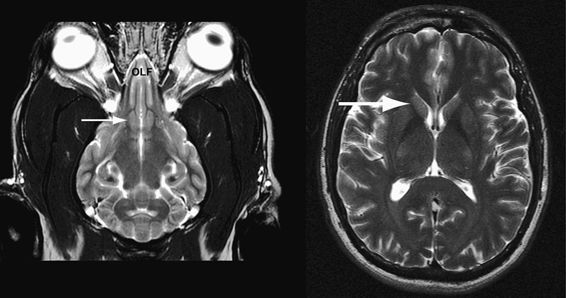
Dorsal plane view of the dog brain showing the olfactory bulb (left) and the corresponding view of the human brain (right). The arrows point to the caudate in both brains.
(Dog brain image by permission of Thomas Fletcher, University of Minnesota; human brain by Gregory Berns)
But what would that tell us?
The whole of the Dog Project hinged on the promise of figuring out what dogs think. Even if we succeeded in finding the parts of the brain that responded to different hand signals, that wouldn’t necessarily mean that we knew what the dogs were thinking. To answer this deeper question, we would have to interpret the patterns of activation based on similar patterns in humans. If we saw activity in parts of the dog brain that we could identify, and we knew what those parts did in humans, we could begin to build a functional map of the canine brain. Using the concept of homology, we could infer canine thought processes from their human equivalents.
This was a shaky premise.
In recent years, there has been a bit of a scientific backlash against neuroimaging. Functional MRI has made it easy to dream up poorly controlled experiments and have groups of undergraduates go into the scanner. Many scientists, eager to get a quick publication in a high-profile journal, overinterpreted the patterns of activity they found in the human brain. It became commonplace to point to activity in a particular brain region and interpret that as evidence for a particular emotion or other cognitive function. It was too easy to observe activation of a structure and conclude, for example, that the person was feeling happy or sad or fearful or some other emotional state based on the scientist’s assumptions of what different brain regions did. Eventually, neuroscientists termed this type of reasoning reverse inference, and it became a key factor in rejecting many fMRI papers.
I had always felt that the criticism of reverse inference, usually uttered with the same contempt one would have for a bag of doo-doo, was overblown. I wouldn’t fault scientists for overinterpreting their data. If I doubted their conclusions, I could always look at their results and draw my own inferences. If I didn’t believe their results, I wouldn’t cite them in my papers. Good and valid conclusions stand the test of time, while false ones fade into obscurity and are eventually forgotten.
The Dog Project would not only be relying on reverse inference, it would depend on reverse inference of a dog’s brain as if it were a human’s. Interspecies reverse inference. I could already imagine what my colleagues would say about this.
Fortunately, Andrew and I had decided to stick with what we knew—the reward system. Our task of deciphering function in the dog brain was going to be a lot easier. Unlike the cortex, with its labyrinthine folds, the reward system belongs to the evolutionarily older reptilian part of the brain. The heart of the reward system is the caudate. Because it is so ancient, all mammals have a caudate, and lucky for us, it looks pretty much the same in dogs and humans.
While neuroscientists can quibble about reverse inference in the cortex, when we did an analysis of reverse inference in the caudate, we found that activity in this region is almost always associated with the expectation of something good. As long as we stuck to the caudate, we would be safe in interpreting activity in this part of the dog’s brain as being a signal of a positive feeling. Everything else we found would have to be interpreted with caution.
Even if we limited ourselves to simple questions of whether the dog had positive feelings based on caudate activation, we could still accomplish a lot with brain imaging. No longer would we be stuck interpreting dogs’ behavior based on tail wagging, which is an imperfect indicator of the emotional state of a dog. Dogs wag their tails when they’re happy, when they’re anxious, or when they’re unsure of what else to do. I still wanted to know if our dogs reciprocated our love for them in any way. And although love is a complicated human emotion, the positive aspects of it have been consistently associated with caudate activation.
The first experiment was a proof of concept. Before we could move on to complicated questions, like love, we first had to demonstrate that we could measure caudate activity in the dog. But that wouldn’t be enough. We would have to show that we could interpret that activity in terms of how much the dogs liked something. Because hot dogs are so much better than peas, especially to a dog, the hand signal for hot dogs should cause more caudate activity than the signal for peas.